Home
Radiology
Advanced imaging services featuring modern MRI suites, multi-slice CT, digital X-rays, and high-frequency ultrasound. Expert radiologist consultation included.
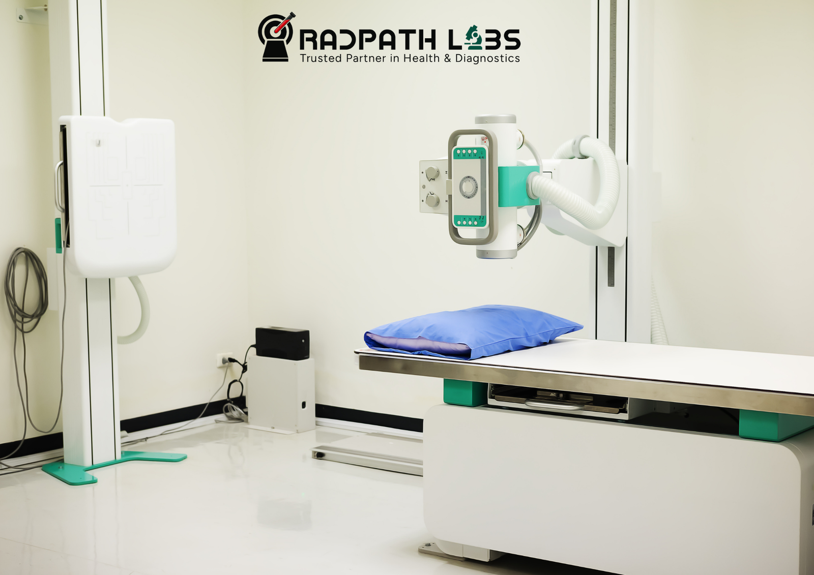
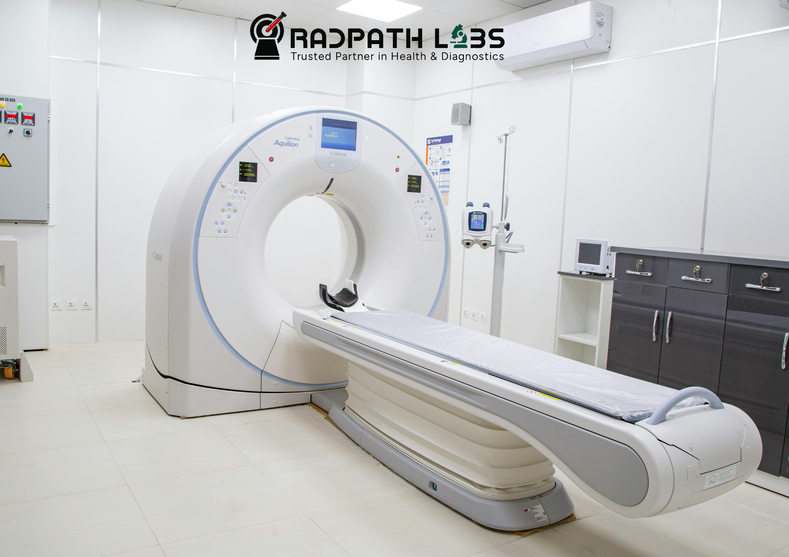
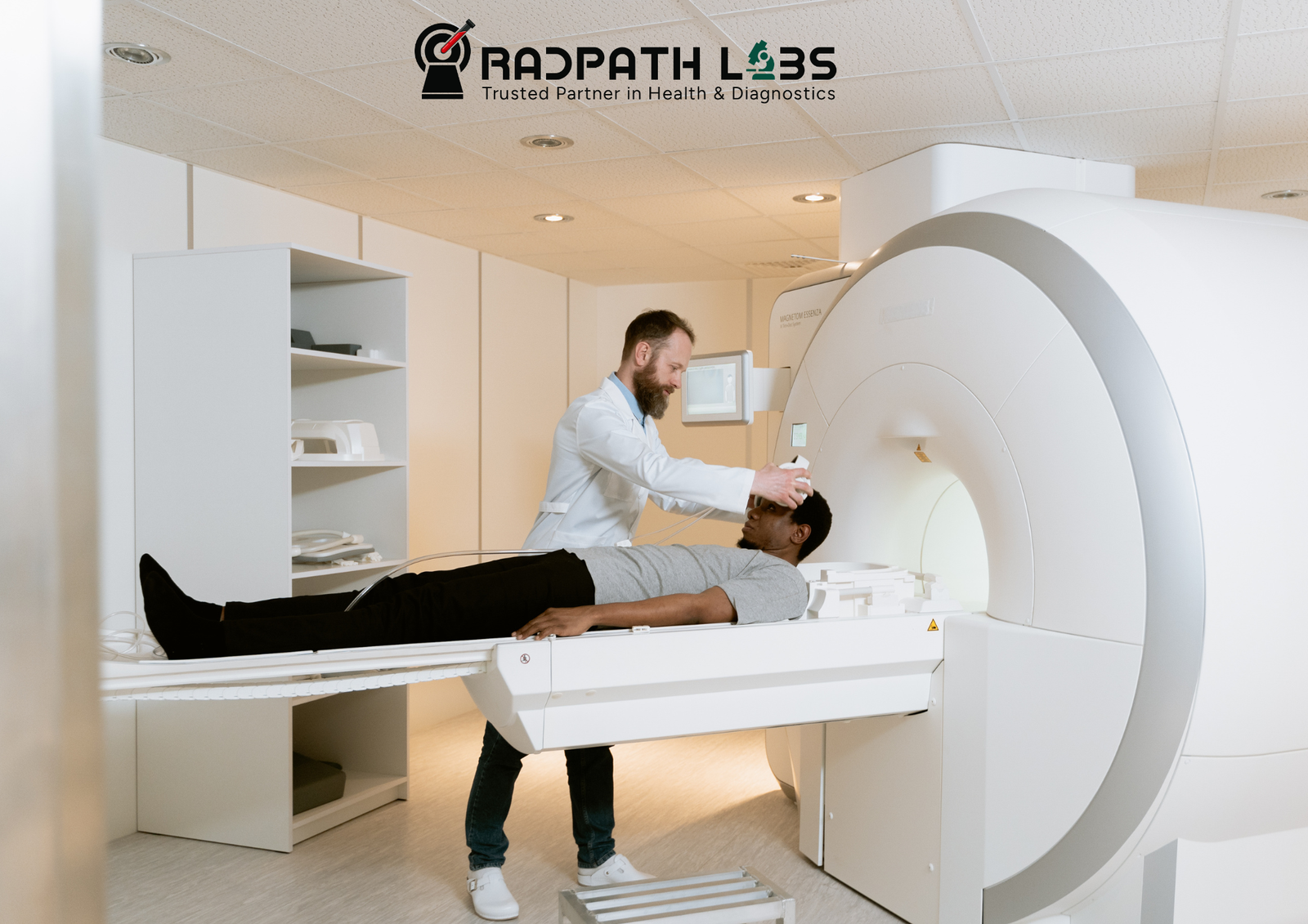
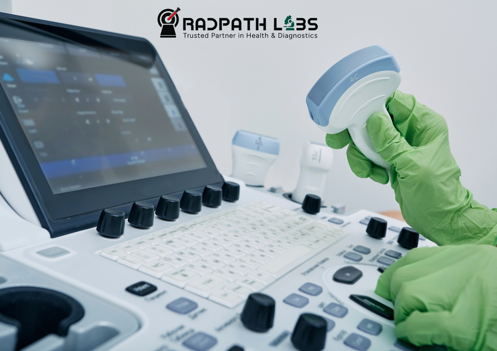
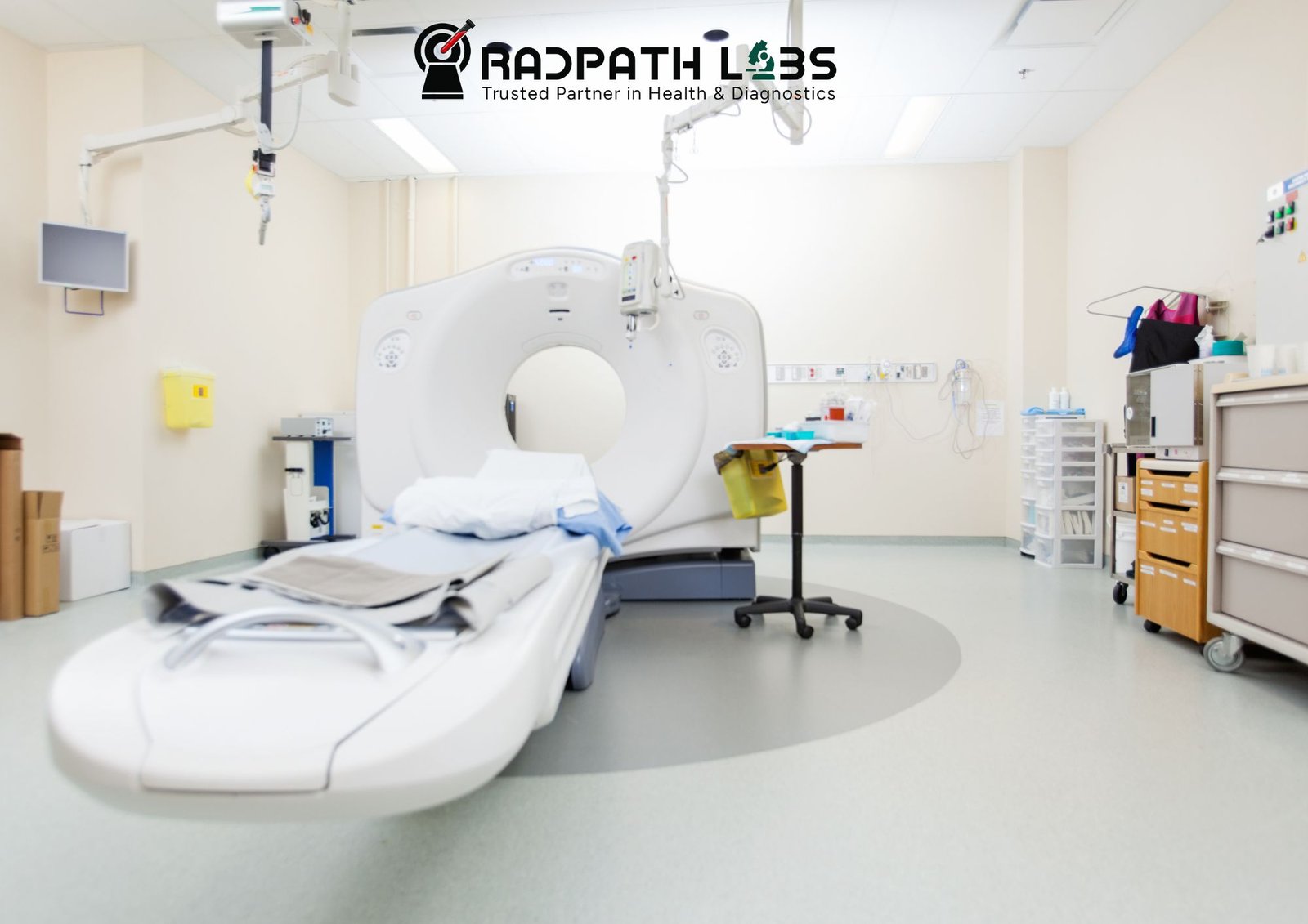
Advanced imaging services featuring modern MRI suites, multi-slice CT, digital X-rays, and high-frequency ultrasound. Expert radiologist consultation included.
